It is the continuation of the deep branch of the radial nerve after this has crossed the supinator muscleIt is considerably diminished in size compared to the deep branch of the radial nerveThe nerve fibers originate from cervical segments C7 and C8 in the spinal column. It is the surface of the body opposite from the chest and the abdomenThe vertebral column runs the length of the back and creates a central area of recession.
![]()
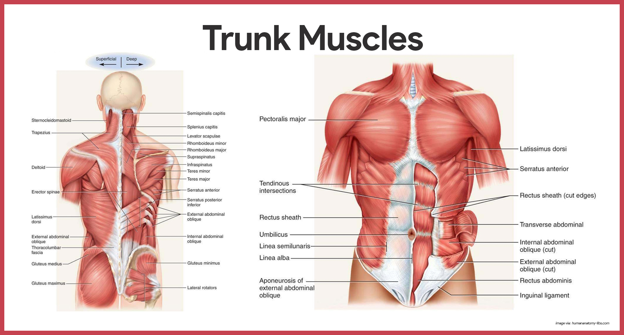
Muscles Of The Trunk Anatomy Diagram Pictures Kenhub
The deep back muscles also called intrinsic or true back muscles consist of four layers of muscles.
. Refers to parts of the body away from the trunk or center of the body. Overview of the Muscular System. The posterior interosseous nerve or dorsal interosseous nerve is a nerve in the forearm.
The breadth of the back is created by the shoulders at the top and the pelvis. The distal bile duct is the farthest end of the cystic duct away from the gallbladder. Proximo near-al pertaining to.
The human back also called the dorsum is the large posterior area of the human body rising from the top of the buttocks to the back of the neck. Please help BlueLink grow by filling out this 2 minute survey to help us understand our users. The second migratory pathway takes the trunk neural crest cells ventrolaterally through the anterior half of each sclerotome.
The trunk neural crest whose cells take one of two major pathwaysNeural crest cells that become the pigment-synthesizing melanocytes migrate dorsolaterally into the ectoderm and continue on their way toward the ventral midline of the belly. The hamstrings are closely related to each. Posterior thigh muscles Hamstring muscles The hamstring muscles or simply the hamstrings are a group of three long muscles located in the posterior compartment of the thigh shaping up the surface anatomy of this regionThese muscles are the biceps femoris semimembranosus and semitendinosus muscles.
For example the wrist joint is distal to the elbow. Deep back muscles. On the anterior and posterior views of the muscular system above superficial muscles those at the surface are shown on the right side of the body while deep muscles those underneath the superficial muscles are shown on the left half of the body.
Superficial intermediate deep and deepest layersThese muscles lie on each side of the vertebral column deep to the thoracolumbar fasciaThey span the entire length of the vertebral column extending from the cranium to the pelvis. Denoting tail or hindposterior part of the body.

Muscles Of The Trunk Posterior View Illustration By Alan Gesek Medical Illustration Animation

Posterior Trunk Muscles Diagram Quizlet

Muscles Of The Posterior Trunk Quiz
Muscular System Anatomy And Physiology Nurseslabs

Muscles Of Posterior Trunk Labeling Diagram Quizlet

File 79201024006 Posterior Trunk Muscles Posterior Trunk Muscles Trunk Muscles Muscles Of The Trunk Diagram Human Body Anatomy Human Anatomy Muscle Anatomy
![]()
Muscles Of The Trunk Anatomy Diagram Pictures Kenhub

Posterior Trunk Muscles Diagram Quizlet
0 comments
Post a Comment